Esoma-KE Logo
Change Class  |  Change Subject

Gaseous Exchange in Animals - High school Biology

  Change Class FORM ONE
Select Subject  |  Biology

BIOLOGY REVISION QUESTIONS

Click to Attempt Form 2 Biology Questions


Want to enjoy learning? Try reading without Ads!

Say goodbye to distractions and enjoy uninterrupted learning with our new ad-free feature. Dive into our vast collection of educational content without any interruptions.

Explore Ad-Free learning plus lots of other premium benefits for as little as KES 200 per month.


Last Updated: 31-12-2021 | Esoma-KE

Gaseous Exchange in Animals

- All animals take in oxygen for oxidation of organic compounds to provide energy for cellular activities.
- The carbon (IV) oxide produced as a by-product is harmful to cells and has to be constantly removed from the body.
- Most animals have structures that are adapted for taking in oxygen and for removal of carbon (IV) oxide from the body.
- These are called respiratory organs.
- The process of taking in oxygen into the body and carbon (IV) oxide out of the body is called breathing or ventilation.
- Gaseous exchange involves passage of oxygen and carbon (IV) oxide through a respiratory surface by diffusion.

Types and Characteristics of Respiratory surfaces

- Different animals have different respiratory surfaces.
- The type depends mainly on the habitat of the animal, size, shape and whether body form is complex or simple.
- Cell Membrane: In unicellular organisms the cell membrane serves as a respiratory surface.
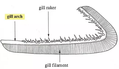
- Gills: Some aquatic animals have gills which may be external as in the tadpole or internal as in bony fish e.g. tilapia.
- They are adapted for gaseous exchange in water.
- Skin: Animals such as earthworm and tapeworm use the skin or body surface for gaseous exchange.
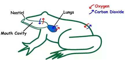
- The skin of the frog is adapted for gaseous exchange both in water and on land.
- The frog also uses epithelium lining of the mouth or buccal cavity for gaseous exchange.
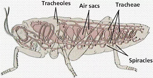
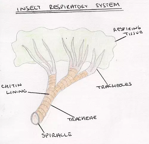
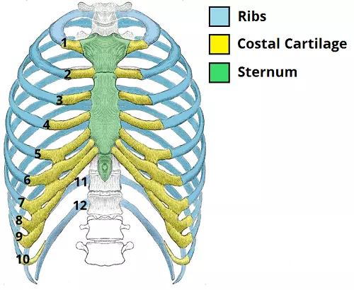
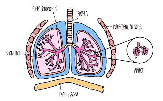
- Lungs: Mammals, birds and reptiles have lungs which are adapted for gaseous exchange.

Characteristics of Respiratory Surfaces

- They are permeable to allow entry of gases.
- They have a large surface area in order to increase diffusion.
- They are usually thin in order to reduce the distance of diffusion.
- They are moist to allow gases to dissolve.
- They are well-supplied with blood to transport gases and maintain a concentration gradient.


Gaseous Exchange in Amoeba

- Gaseous exchange occurs across the cell membrane by diffusion.
- Oxygen diffuses in and carbon (IV) oxide diffuses out.
- Oxygen is used in the cell for respiration making its concentration lower than that in the surrounding water.
- Hence oxygen continually enters the cell along a concentration gradient.
- Carbon (IV) oxide concentration inside the cell is higher than that in the surrounding water thus it continually diffuses out of the cell along a concentration gradient.

Gaseous Exchange in Insects

- Gaseous exchange in insects e.g., grasshopper takes place across a system of tubes penetrating into the body known as the tracheal system.

Grasshopper's Respiratory System - biology Form two
Grasshopper's Respiratory System - Image Courtesy

Insect's Respiratory System - biology Form two
Insect's Respiratory System - Image Courtesy

- The main trachea communicate with atmosphere through tiny pores called spiracles.
- Spiracles are located at the sides of body segments;
- Two pairs on the thoracic segments and eight pairs on the sides of abdominal segments.
- Each spiracle lies in a cavity from which the trachea arises.
- Spiracles are guarded with valves that close and thus prevent excessive loss of water vapour.
- A filtering apparatus i.e. hairs also traps dust and parasites which would clog the trachea if they gained entry.
- The valves are operated by action of paired muscles.


Mechanism of Gaseous Exchange in Insects

- The main tracheae in the locust are located laterally along the length of the body on each side and they are interconnected across.
- Each main trachea divides to form smaller tracheae, each of which branches into tiny tubes called tracheoles.
- Each tracheole branches further to form a network that penetrates the tissues. Some tracheoles penetrate into cells in active tissue such as flight muscles.
- These are referred to as intracellular tracheoles.
- Tracheoles in between the cells are known as intercellular tracheoles.
- The main tracheae are strengthened with rings of cuticle.
- This helps them to remain open during expiration when air pressure is low.

Adaptation of Insect Tracheoles for Gaseous Exchange

- The fine tracheoles are very thin about one micron in diameter in order to permeate tissue.
- They are made up of a single epithelial layer and have no spiral thickening to allow diffusion of gases.
- Terminal ends of the fine tracheoles are filled with a fluid in which gases dissolve to allow diffusion of oxygen into the cells.
- Amount of fluid at the ends of fine tracheoles varies according to activity i.e. oxygen demand of the insect.
- During flight, some of the fluid is withdrawn from the tracheoles such that oxygen reaches muscle cells faster and the rate of respiration is increased.
- In some insects, tracheoles widen at certain places to form air sacs.
- These are inflated or deflated to facilitate gaseous exchange as need arises.
- Atmospheric air that dissolves in the fluid at the end of tracheoles has more oxygen than the surrounding cells of tracheole epithelium.
- Oxygen diffuses into these cells along a concentration gradient.
- Carbon (IV) oxide concentration inside the cells is higher than in the atmospheric.
- Air and diffuses out of the cells along a concentration gradient.
- It is then removed with expired air.


Ventilation in Insects

- Ventilation in insects is brought about by the contraction and relaxation of the abdominal muscles.
- In locusts, air is drawn into the body through the thoracic spiracles and expelled through the abdominal spiracles.
- Air enters and leaves the tracheae as abdominal muscles contract and relax.
- The muscles contract laterally so the abdomen becomes wider and when they relax it becomes narrow.
- Relaxation of muscles results in low pressure hence inspiration occurs while contraction of muscles results in higher air pressure and expiration occurs.
- In locusts, air enters through spiracles in the thorax during inspiration and leaves through the abdominal spiracles during expiration.
- This results in efficient ventilation.
- Maximum extraction of oxygen from the air occurs sometimes when all spiracles close and hence contraction of abdominal muscles results in air circulating within the tracheoles.
- The valves in the spiracles regulate the opening and closing of spiracles.
Observation of Spiracle in Locust
- Some fresh grass is placed in a gas jar.
- A locust is introduced into the jar.
- A wire mesh is placed on top or muslin cloth tied around the mouth of the beaker with rubber band.
- The insect is left to settle.
- Students can approach and observe in silence the spiracles and the abdominal movements during breathing.
- Alternatively the locust is held by the legs and observation of spiracles is made by the aid of hand lens.

Gaseous Exchange in Bony Fish (e.g, Tilapia)

- Gaseous exchange in fish takes place between the gills and the surrounding water.
- The gills are located in an opercular cavity covered by a flap of skin called the operculum.
- Each gill consists of a number of thin leaf-like lamellae projecting from a skeletal base branchial arch (gill bar) situated in the wall of the pharynx.
- There are four gills within the opercular cavity on each side of the head.
- Each gill is made up of a bony gill arch which has a concave surface facing the mouth cavity (anterior) and a convex posterior surface.
- Gill rakers are bony projections on the concave side that trap food and other solid particles which are swallowed instead of going over and damaging the gill filaments.
- Two rows of gill filaments subtend from the convex surface.

Adaptation of Gills for Gaseous Exchange

- Gill filaments are thin walled.
- Gill filaments are very many (about seventy pairs on each gill), to increase surface area.
- Each gill filament has very many gill lamellae that further increase surface area.
- The gill filaments are served by a dense network of blood vessels that ensure efficient transport of gases.
- It also ensures that a favourable diffusion gradient is maintained.
- The direction of flow of blood in the gill lamellae is in the opposite direction to that of the water (counter current flow) to ensure maximum diffusion of gases.

A Fish Gill - biology Form two
A Fish Gill - Image Courtesy

Ventilation

- As the fish opens the mouth, the floor of the mouth is lowered.
- This increases the volume of the buccal cavity.
- Pressure inside the mouth is lowered causing water to be drawn into the buccal cavity.
- Meanwhile, the operculum is closed, preventing water from entering or leaving through the opening.
- As the mouth closes and the floor of the mouth is raised, the volume of buccal cavity decreases while pressure in the opercular cavity increases due to contraction of opercular muscles.
- The operculum is forced to open and water escapes.
- As water passes over the gills, oxygen is absorbed and carbon dioxide from the gills dissolves in the water.
- As the water flows over the gill filaments oxygen in the water is at a higher concentration than that in the blood flowing, in the gill.
- Oxygen diffuses through the thin walls of gill filaments/lamellae into the blood.
- Carbon (IV) oxide is at a higher concentration in the blood than in the water.
- It diffuses out of blood through walls of gill filaments into the water.

Counter Current Flow

- In the bony fish direction of flow of water over the gills is opposite that of blood flow through the gill filaments.
- This adaptation ensures that maximum amount of oxygen diffuses from the water into the blood in the gill filament.
- This ensures efficient uptake of oxygen from the water.
- Where the flow is along the same direction (parallel flow) less oxygen is extracted from the water.


Gaseous Exchange in an Amphibian - Frog

- An adult frog lives on land but goes back into the water during the breeding season.
- A frog uses three different respiratory surfaces.
- These are the skin, buccal cavity and lungs.

Gaseous Exchange in a Frog - biology Form two
Image Courtesy

Skin

- The skin is used both in water and on land.
- It is quite efficient and accounts for 60% of the oxygen taken in while on land.

Adaptations of a Frog's Skin for Gaseous Exchange

- The skin is a thin epithelium to allow fast diffusion.
- The skin between the digits in the limbs (i.e. webbed feet) increase the surface area for gaseous exchange.
- It is richly supplied with blood vessels for transport of respiratory gases.
- The skin is kept moist by secretions from mucus glands.
- This allows for respiratory gases to dissolve.
- Oxygen dissolved in the film of moisture diffuses across the thin epithelium and into the blood which has a lower concentration of oxygen.
- Carbon (IV) oxide diffuses from the blood across the skin to the atmosphere along the concentration gradient.

Buccal (Mouth) Cavity

- Gaseous exchange takes place all the time across thin epithelium lining the mouth cavity.
- Adaptations of Buccal Cavity for Gaseous Exchange.
- It has a thin epithelium lining the walls of the mouth cavity allowing fast diffusion of gases.
- It is kept moist by secretions from the epithelium for dissolving respiratory gases.
- It has a rich supply of blood vessels for efficient transport of respiratory gases.
- The concentration of oxygen in the air within the mouth cavity is higher than that of the blood inside the blood vessels.
- Oxygen, therefore dissolves in the moisture lining the mouth cavity and then diffuses into the blood through the thin epithelium.
- On the other hand, carbon (IV) oxide diffuses in the opposite direction along a concentration gradient.

Lungs

- There is a pair of small lungs used for gaseous exchange.

Adaptation of Lungs

- The lungs are thin walled for fast diffusion of gases.
- Have internal foldings to increase surface area for gaseous exchange.
- A rich supply of blood capillaries for efficient transport of gases.
- Moisture lining for gases to dissolve.

Ventilation

Inspiration

- During inspiration, the floor of the mouth is lowered and air is drawn in through the nostrils.
- When the nostrils are closed and the floor of the mouth is raised, air is forced into the lungs.
- Gaseous exchange occurs in the lungs, oxygen dissolves in the moisture lining of the lung and diffuses into the blood through the thin walls.
- Carbon (IV) oxide diffuses from blood into the lung lumen.

Expiration

- When the nostrils are closed and the floor of mouth is lowered by contraction of its muscles, volume of mouth cavity increases.
- Abdominal organs press against the lungs and force air out of the lungs into buccal cavity.
- Nostrils open and floor of the mouth is raised as its muscles relax.
- Air is forced out through the nostrils.


Gaseous Exchange in Lungs - Human

- The breathing system of a mammal consists of a pair of lungs which are thin-walled elastic sacs lying in the thoracic cavity.
- The thoracic cavity consists of vertebrae, sternum, ribs and intercostal muscles.

vertebrae, sternum, ribs and intercostal muscles - biology Form two
Vertebrae, Sternum, Ribs and Intercostal Muscles - Image Courtesy of Teach Me Anatomy

- The thoracic cavity is separated from the abdominal cavity by the diaphragm.
- The lungs lie within the thoracic cavity.
- They are enclosed and protected by the ribs which are attached to the sternum and the thoracic vertebrae.
- There are twelve pairs of ribs, the last two pairs are called 'floating ribs' because they are only attached to the vertebral column.
- The ribs are attached to and covered by internal and external intercostals muscles.
- The diaphragm at the floor of thoracic cavity consists of a muscLe sheet at the periphery and a central circular fibrous tissue.
- The muscles of the diaphragm are attached to the thorax wall.
- The lungs communicate with the outside atmosphere through the bronchi, trachea, mouth and nasal cavities.
- The trachea opens into the mouth cavity through the larynx.
- A flap of muscles, the epiglottis, covers the opening into the trachea during swallowing.
- This prevents entry of food into the trachea.
- Nasal cavities are connected to the atmosphere through the external nares(or nostrils)which are lined with hairs and mucus that trap dust particles and bacteria, preventing them from entering into the lungs.
- Nasal cavities are lined with cilia.
- The mucus traps dust particles,
- The cilia move the mucus up and out of the nasal cavities.
- The mucus moistens air as it enters the nostrils.
- Nasal cavities are winding and have many blood capillaries to increase surface area to ensure that the air is warmed as it passes along.

Lungs Diagram

Human Respiratory System - biology Form two
Human Respiratory System - Image Courtesy of The Science Hive

- Each lung is surrounded by a space called the pleural cavity.
- It allows for the changes in lung volume during breathing.
- An internal pleural membrane covers the outside of each lung while an external pleural membrane lines the thoracic wall.
- The pleural membranes secrete pleural fluid into the pleural cavity.
- This fluid prevents friction between the lungs and the thoracic wall during breathing.
- The trachea divides into two bronchi, each of which enters into each lung.
- Trachea and bronchi are lined with rings of cartilage that prevent them from collapsing when air pressure is low.
- Each bronchus divides into smaller tubes, the bronchioles.
- Each bronchiole subdivides repeatedly into smaller tubes ending with fine bronchioles.
- The fine bronchioles end in alveolar sacs, each of which gives rise to many alveoli.
- Epithelium lining the inside of the trachea, bronchi and bronchioles has cilia and secretes mucus.


Adaptations of Alveolus to Gaseous Exchange

- Each alveolus is surrounded by very many blood capillaries for efficient transport of respiratory gases.
- There are very many alveoli that greatly increases the surface area for gaseous exchange.
- The alveolus is thin walled for faster diffusion of respiratory gases.
- The epithelium is moist for gases to dissolve.

Gaseous Exchange Between the Alveoli and the Capillaries

- The walls of the alveoli and the capillaries are very thin and very close to each other.
- Blood from the tissues has a high concentration of carbon (IV) oxide and very little oxygen compared to alveolar air.
- The concentration gradient favours diffusion of carbon (IV) oxide into the alveolus and oxygen into the capillaries.
- No gaseous exchange takes place in the trachea and bronchi.
- These are referred to as dead space.

Ventilation

- Exchange of air between the lungs and the outside is made possible by changes in the volumes of the thoracic cavity.
- This volume is altered by the movement of the intercostal muscles and the diaphragm.

Inspiration

- The ribs are raised upwards and outwards by the contraction of the external intercostal muscles, accompanied by the relaxation of internal intercostal muscles.
- The diaphragm muscles contract and diaphragm moves downwards.
- The volume of thoracic cavity increases, thus reducing the pressure.
- Air rushes into the lungs from outside through the nostrils.

Expiration

- The internal intercostal muscles contract while external ones relax and the ribs move downwards and inwards.
- The diaphragm muscles relaxes and it is pushed upwards by the abdominal organs. It thus assumes a dome shape.
- The volume of the thoracic cavity decreases, thus increasing the pressure.
- Air is forced out of the lungs.
- As a result of gaseous exchange in the alveolus, expired air has different volumes of atmospheric gases as compared to inspired air.

Comparison of Inspired and Expired Air (% by volume)

Component Inspired Expired %
Oxygen 21 16
Carbon 0.03 4
Nitrogen 79 79
Moisture Variable Saturated

Lung Capacity

- The amount of air that human lungs can hold is known as lung capacity.
- The lungs of an adult human are capable of holding 5,000 cm3 of air when fully inflated.
- However, during normal breathing only about 500 cm3 of air is exchanged.
- This is known as the tidal volume.
- A small amount of air always remains in the lungs even after a forced expiration.
- This is known as the residual volume.
- The volume of air inspired or expired during forced breathing is called vital capacity.

Control of Rate Of Breathing

- The rate of breathing is controlled by the respiratory centre in the medulla of the brain.
- This centre sends impulses to the diaphragm through the phrenic nerve.
- Impulses are also sent to the intercostal muscles.
- The respiratory centre responds to the amount of carbon (IV) oxide in the blood.
- If the amount of carbon (IV) oxide rises, the respiratory centre sends impulses to the diaphragm and the intercostal muscles which respond by contracting in order to increase the ventilation rate.
- Carbon (IV) oxide is therefore removed at a faster rate.

Factors Affecting Rate of Breathing in Humans

- Factors that cause a decrease or increase in energy demand directly affect rate of breathing.
- Exercise, any muscular activity like digging.
- Sickness.
- Emotions like anger, flight.
- Sleep.

Effects of Exercise on Rate of Breathing

- Students to work in pairs.
- One student stands still while the other counts (his/her) the number of breaths per minute.
- The student whose breath has been taken runs on the sport vigorously for 10 minutes.
- At the end of 10 minutes the number of breaths per minute is immediately counted and recorded.
- It is noticed that the rate of breathing is much higher after exercise than at rest.


Diseases of the Respiratory System

1. Asthma

- Asthma is a chronic disease characterised by narrowing of air passages.

Causes:

a) Allergy
- Due to pollen, dust, fur, animal hair, spores among others.
- If these substances are inhaled, they trigger release of chemical substances and they may cause swelling of the bronchioles and bring about an asthma attack.
b) Heredity
- Asthma is usually associated with certain disorders which tend to occur in more than one member of a given family, thus suggesting' a hereditary tendency.
c) Emotional or mental stress
- Strains the body immune system hence predisposes to asthma attack.

Symptoms

- Asthma is characterized by wheezing and difficulty in breathing accompanied by feeling of tightness in the chest as a result of contraction of the smooth muscles lining the air passages.

Treatment and Control

- There is no definite cure for asthma.
- The best way where applicable is to avoid whatever triggers an attack (allergen).
- Treatment is usually by administering drugs called bronchodilators.
- The drugs are inhaled, taken orally or injected intravenously depending on severity of attack to relief bronchial spasms.

2. Bronchitis

- This is an inflammation of bronchial tubes.

Causes

- This is due to an infection of bronchi and bronchioles by bacteria and viruses.

Symptoms

- Difficulty in breathing.
- Cough that produces mucus.

Treatment

- Antibiotics are administered.

3. Pulmonary Tuberculosis

- Tuberculosis is a contagious disease that results in destruction of the lung tissue.

Causes

- Tuberculosis is caused by the bacterium Mycobacterium tuberculosis.
- Human tuberculosis is spread through droplet infection i.e., in saliva and sputum.
- Tuberculosis can also spread from cattle to man through contaminated milk.
- From a mother suffering from the disease to a baby through breast feeding.
- The disease is currently on the rise due to the lowered immunity in persons with HIV and AIDS (Human Immuno Deficiency Syndrome).
- Tuberculosis is common in areas where there is dirt, overcrowding and malnourishment.

Symptoms

- It is characterised by a dry cough, lack of breath and body wasting.

Prevention

- Proper nutrition with a diet rich in proteins and vitamins to boost immunity.
- Isolation of sick persons reduces its spread.
- Utensils used by the sick should be sterilised by boiling.
- Avoidance of crowded places and living in well ventilated houses.
- Immunisation with B.C.G. vaccine gives protection against tuberculosis.
- This is done a few days after birth with subsequent boosters.

Treatment

- Treatment is by use of antibiotics.

4. Pneumonia

- Pneumonia is infection resulting in inflammation of lungs.
- The alveoli get filled with fluid and bacterial cells decreasing surface are for gaseous exchange.
- Pneumonia is caused by bacteria and virus.
- More infections occur during cold weather.
- The old and the weak in health are most vulnerable.

Symptoms

- Pain in the chest accompanied by a fever, high body temperatures (39-40”ĘC) and general body weakness.

Prevention

- Maintain good health through proper feeding.
- Avoid extreme cold.

Treatment

- If the condition is caused by pneumococcus bacteria, antibiotics are administered.
- If breathing is difficult, oxygen may be given using an oxygen mask.

5. Whooping Cough

- Whooping cough is an acute infection of respiratory tract.
- The disease is more common in children under the age of five but adults may also be affected.

Causes

- It is caused by Bordetella pertusis bacteria and is usually spread by droplets produced when a sick person coughs.

Symptoms:

- Severe coughing and frequent vomiting.
- Thick sticky mucus is produced.
- Severe broncho-pneumonia.
- Convulsions in some cases.

Prevention

- Children may be immunised against whooping cough by means of a vaccine which is usually combined with those against diphtheria and tetanus.
- It is called Triple Vaccine or Diptheria, Pertusis and Tetanus (DPT).

Treatment

- Antibiotics are administered.
- To reduce the coughing, the patient should be given drugs.


Gaseous Exchange in Animals Quiz

Question - of -


Hello Esoma-KE Member, please help review these notes.    Why review

Your review has been successfully submitted.
Tell us what you think about the notes.

Submit Comment

Students' and Teachers' Reviews


Here's what our users had to say ...

No comments given yet! Be the first to comment.