Female Reproduction System
Female Reproductive System Diagram
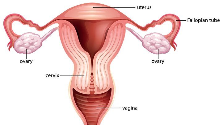
(Image Courtesy)
The female reproduction system consist of the following:
a) Ovaries
- Are two oval cream coloured structures found in lower abdomen below the kidneys.
b) Oviducts
- They produce the ova.
- Are tubes which conduct the ova produced by the ovaries to the uterus.
- Fertilization occurs in the upper part of the oviduct.
c) Uterus
- The uterus is a hollow muscular organ found in the lower abdomen.
- The embryo develops inside the uterus.
- The inner lining endometrium supplies nutrients to embryo.
- The embryo is implanted into the inner uterine wall- the endometrium which nourishes the embryo.
- The thick muscles of the uterus assist in parturition.
d) Cervix
- Has a ring of muscles that separates the uterus from the vagina.
- It forms the opening to the uterus.
e) Vagina
- Is a tube that opens to the outside and it acts as the copulatory and birth canal through the vulva.
Male Reproductive System
Male Reproductive System Diagram
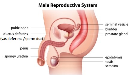
(Image Courtesy)
The male reproductive system consists of the following:
a) Testis
- Each testis is a mass of numerous coiled tubes called seminiferous tubules.
- Each is enclosed within a scrotal sac that suspends them between the thighs.
- This ensures that sperms are maintained at a temperature lower than that of the main body.
b) Seminiferous tubules
- The lining of seminiferous tubules consists of actively dividing cells which give rise to sperms.
- Between the seminiferous tubules are interstitial cells which produce the male hormones called androgens e.g. testosterone.
- The seminiferous tubules unite to form the epididymis, which is a coiled tube where sperms are stored temporarily.
- Vas deferens (sperm duct) is the tube through which sperms are carried from testis to urethra.
- Seminal vesicle produces an alkaline secretion which nourishes the spermatozoa.
c) Prostate gland
- Produces an alkaline secretion to neutralise vaginal fluids.
d) Cowper's' gland
- Secretes an alkaline fluid.
- All these fluids together with spermatozoa form semen.
e) Urethra
- Is a long tube through which the semen is conducted during copulation.
- It also removes urine from the bladder.
f) Penis
- Is an intromittent organ which is inserted into the vagina during copulation.
Fertilization in Animals
- Fertilization is preceded by copulation in which the erect penis is inserted into the vagina.
- This leads to ejaculation of semen.
- The sperms swim through the female's genital tract to the upper part of the oviduct.
- The head of the sperm penetrates the egg after the acrosome_ releases lytic enzymes t dissolve the egg membrane.
- The tail is left behind.
- Sperm nucleus fuses with that of the ovum and a zygote is formed.
- A fertilization membrane forms around the zygote which prevents other sperms from penetrating the zygote.
Implantation
- After fertilization the zygote begins to divide mitotically as it moves towards the uterus.
- It becomes embedded in the wall of the uterus a process called implantation.
- By this time the zygote is a hollow ball of cells called blastocyst or embryo.
- In the uterus the embryo develops villi which project into uterus for nourishment later the villi and endometrium develop into placenta.
Embryonic Membranes
- Embryonic membranes develop around the embryo.
- The outermost membrane is the chorion which forms the finger-like projections (chorionic villi) which supply nutrients to the embryo.
- The amnion surrounds the embryo forming a fluid filled cavity within which the embryo lies.
- Amniotic cavity is filled with amniotic fluid.
- This fluid acts as a shock absorber and protects the foetus against mechanical injury.
- It also regulates temperature.
- The chorionic villi, allantois together with the endometrium from the placenta.
- The embryo is attached to the placenta by a tube called umbilical cord which has umbilical vein and artery.
- The maternal blood in the placenta flows in the spaces lacuna and surrounds capillaries from umbilical vein and artery.
- The umbilical cord increase in length as the embryo develops.
Role of Placenta
1) Protection
- Maternal blood and foetal blood do not mix.
- This ensures that the pathogens and toxins from maternal blood do not reach the foetus.
- The placenta allows maternal antibodies to pass into the foetus, providing the foetus with immunity.
2) Nutrition
- The placenta facilitates the transfer of nutrients from maternal blood to foetus.
3) Excretion
- Placenta facilitates the removal of nitrogenous wastes from the foetus' blood to maternal blood.
4) Gaseous exchange
- Oxygen from the maternal blood diffuses into the foetal blood while carbon (IV) oxide from foetal blood diffuse into maternal blood.
5) Production of hormones
- Placenta produces progesterone and oestrogen.
Gestation period
- The period between conception and birth is called gestation.
- In humans gestation takes nine months (40 weeks).
- The embryo differentiates into tissues and organs during this period.
Week 1 to 3:
- Zygote divides to form blastocyst.
- Implantation takes place.
- The three germ layers form endoderm, mesoderm and ectoderm.
- Nervous system starts to form.
Week 4 to 7:
- Development of circulating and digestive systems.
- Further development of nervous system, formation of sensory organs.
- All major internal organs are developed.
- At week 5, heartbeat starts.
Week 8 to 24:
- All organs well developed including sex organs.
- Hair, finger and toe nails grow.
- Foetus move and eyelids open.
Week 25- 30:
- The fully developed foetus responds to touch and noises and moves vigorously.
- The head turns and faces downwards ready for birth.
Week 31-40:
- Foetus increases in size.
- Birth occurs.
Sources of Hormones and their Function
| Hormone |
Source |
Functions |
| Follicle Stimulating Hormone (FSH) |
Pituitary gland |
Development of ovarian follicle; stimulates secretion of oestrogen by the ovary |
| Luteinising Hormone |
Pituitary gland |
Causes ovulation; causes development of Graafian follicle into the corpus luteurn; causes secretion of progesterone by the ovary |
| Prolactin |
Pituitary gland |
Initiates production and secretion of milk by the mammary glands |
| Oxytocin |
Pituitary gland |
Causes contraction of the uterus during parturition (birth) |
| Progesterone |
Corpus luteum in the ovary |
Causes contraction of wall of the uterus to thicken after ovulation |
| Oestrogen |
Ovary |
Causes changes in the uterine wall in preparation for implantation; initiates development of secondary sexual characteristics |
| Androgens-Testosterone |
Interstitial cells of testis |
Stimulates the development of secondary sexual characteristics |
| Interstitial Cell Stimulating Hormone (lCSH) |
Pituitary gland |
Stimulates the interstitial cells of testis to release androgens |
| Human Chorionic Gonadotrophin (HCG) |
Chorionic villi |
Stops the degeneration of the corpus luteum for production of oestrogen and progesterone |
Secondary Sexual Characteristics
Male
- Testosterone is the main androgen that stimulates the development of secondary sexual characteristics.
- Broadening of the shoulders.
- Deepening of the voice due to enlargement of larynx.
- Hair at the pubic area, armpit and chin regions.
- Penis and testis enlarge and produce sperms.
- Body becomes more masculine.
Female
- Enlargement of mammary glands.
- Hair grows around pubic and armpit regions.
- Widening of the hips.
- Ovaries mature and start producing ova.
- Menstruation starts.
- Oestrogen triggers the onset of secondary sexual characteristics.
Menstrual Cycle
- This is characterized by discharge of blood and tissue debris (menses) from the uterus every 28 days.
- This is due to the breakdown of the endometrium which occurs when the level of progesterone falls and the girl starts to menstruate.
- The follicle stimulating hormone (FSH) causes the Graafian follicle to develop and also stimulate the ovary to release oestrogen.
- Oestrogen hormone triggers the onset of secondary sexual characteristics.
- Luteinising hormone (L.H) causes the mature ovum to be released from the Graafian follicle - a process called ovulation.
- After ovulation progesterone hormone is produced.
- After menstruation, the anterior lobe of the pituitary gland starts secreting the follicle stimulating hormone (FS.H) which causes the Graafian follicle to develop in the ovary.
- It also stimulates the ovary tissues to secrete oestrogen.
- Oestrogen brings about the repair and healing of the inner lining of the uterus (endometrium) which had been destroyed during menstruation.
- Oestrogen level stimulates the pituitary gland to produce Luteinising Hormone (L.H).
- This hormone makes the mature Graafian follicle to release the ovum into the funnel of oviduct, a process called ovulation.
- After releasing the ovum, the Graafian follicle changes into a yellow body called corpus luteum.
- The Luteinising hormone stimulates the corpus luteum to secrete a hormone called progesterone which stimulates the thickening and vascularisation of endometrium.
- This prepares the uterine wall for implantation of the blastocyst.
- If fertilization takes place, the level of progesterone increases and thus inhibits FSH from stimulating the maturation of another Graafian follicle.
- If fertilization does not occur, the corpus luteum disintegrates and the level of progesterone goes down.
- The endometrium, sloughs off and menstruation occurs.
Sexually Transmitted Infections (STIs)
Gonorrhea
Causes of Gonorrhea
Gonorrhea is caused by Bacterium
Neisseria gonorrhoeae
Transmission of Gonorrhea
- Sexual contact of an infected partner
- From mother to baby during child birth
- It can also be transmitted through sharing towels although the chances are arguably low. This is beacuse
Neisseria gonorrhoeae cannot survive outside the body for long.
Symptoms of Gonorrhea
- Discharge from the penis or vagina
- Testicular pain in men
- Itching of urethra
- vaginal discharge with odour
Prevention and Control of Gonorrhea
- Avoid indiscriminate sex.
- Treat any infected partner
Syphilis
Causes of Syphilis
Syphilis is caused by Bacterium
Treponema pallidum
Transmission of Syphilis
- Sexual contact of an infected partner
- From mother to baby during child birth
- It can also be transmitted through sharing towels although the chances are arguably low.
Symptoms of Syphilis
- Painless sore on the genitals, rectum or mouth
- Rash
- papules on feet, hands, and genital areas
Prevention and Control of Syphilis
- Treat at primary infection stage
- Avoid indiscriminate sex.
Hepatitis
Causes of Hepatitis
- Hepatitis is caused by the Hepatitis B Virus (HBV)
Transmission of Hepatitis
- Sexual contact with an infected person
- Blood-contaminated needles and syringes
- Body fluids of an infected person enters the body of another.
Symptoms of Hepatitis
- Yellowing of the eyes
- Dark-yellow urine
- Abdominal pain
Prevention and Control of Hepatitis
- Vaccination is recommended as a prevention measure
- Avoid indiscriminate sex
- Use disposable needles and syringes
- Have a strict personal hygiene
HIV and AIDS
Causes of HIV and AIDS
HIV and AIDS is caused by the human immunodeficiency virus
Transmission of HIV and AIDS
- Sexual contact with an infected person,br>
- Sharing needles and syringes with an infected person
- From mother ro child during pregnacy, child birth, or breastfeeding.
Symptoms of HIV and AIDS
- Lesions on skin
- Sore throat and painful mouth sores
- Wasting due to Weight loss
Prevention and Control of HIV and AIDS
- Avoid indiscriminate sex
- Use disposable needles and syringes
RELATED NOTES:
CELL DIVISION: MITOSIS AND MEIOSIS- HIGH SCHOOL BIOLOGY FORM 3
Read on cell division, the different mitosis stages and meiosis stages including Prophase, Metaphase, Anaphase and Telophase. Learn significance of mitosis and meiosis.
SEXUAL REPRODUCTION IN PLANTS - HIGH SCHOOL BIOLOGY FORM 3
Read on sexual reproduction in plants and parts of a flower. Read on pollination, definition of pollination, the types of pollination, and methods of seed dispersal.