


The Circulatory System
- Large and complex animals have circulatory systems that consist of tubes, a transport fluid and a means of pumping the fluid.- Blood is the transport fluid which contains dissolved substances and cells.
- The tubes are blood vessels through which dissolved substances are circulated around the body.
- The heart is the pumping organ which keeps the blood in circulation.
- The types of circulatory system exist in animals: open and closed.
- In an open circulatory system;
- The heart pumps blood into vessels which open into body spaces known as haemocoel.
- Blood comes into contact with tissues.
Closed Circulatory System
- Found in vertebrates and annelids where the blood is confined within blood vessels and does not come into direct contact with tissues.Circulatory System of Insects
- In an insect, there is a tubular heart just above the alimentary canal.
- This heart is suspended in a pericardial cavity by ligaments.
- The heart has five chambers and extends along the thorax and abdomen.
- Blood is pumped forwards into the aorta by waves of contractions in the heart.
- It enters the haemocoel and flows towards the posterior.
- The blood flows back into the heart through openings in each chamber called ostia.
- The ostia have valves which prevent the backflow of blood.
- Blood is not used as a medium for transport of oxygen in insects.
- This is because oxygen is supplied directly to the tissues by the tracheal system.
- The main functions of blood in an insect are to transport nutrients, excretory products and hormones.
Circulatory System of Mammals
- Mammals have a closed circulatory system where a powerful heart pumps blood into arteries.
- The arteries divide into smaller vessels called arterioles.
- Each arteriole divides to form a network of capillaries inside the tissues.
- The capillaries eventually re-unite to form venules, which form larger vessels called veins.
- The veins take the blood back to the heart.
- Blood from the heart goes through the pulmonary artery to the lungs and then back to the heart through pulmonary vein.
- This circulation is called pulmonary circulation.
- Oxygenated blood leaves the heart through the aorta and goes to all the tissues of the body.
- From the tissues, deoxygenated blood flows back to the heart through the vena cava.
- This circulation is called systemic circulation.
- In each complete circulation, the blood flows into the heart twice.
- This is called double circulation.
- Some other animals like fish have a single circulation.
- Blood flows only once through the heart for every complete circuit.
Functions and Structure of the Heart
Diagram of the Structure of the Heart
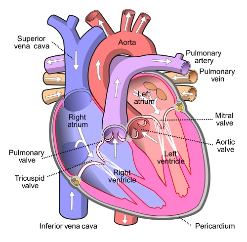
- The heart has four chambers: Two artria (auricles) and two ventricles.
- The left and right side of the heart are separated by a muscle wall (septum) so that oxygenated and deoxygenated blood does not mix.
- Deoxygenated blood from the rest of the body enters the heart through the vena cava.
- Blood enters the right atrium, then through tricuspid valve into right ventricle.
- Then via semi-lunar valve to the pulmonary artery to the lungs.
- Oxygenated blood from the lungs enters the heart through pulmonary vein.
- It enters the left atrium of the heart, then through bicuspid valve into left ventricle.
- Then via semi-lunar valves to aorta which takes oxygenated blood round the body.
- A branch of the aorta called coronary artery supplies blood to the heart muscle.
- The coronary vein carries blood from the heart muscle to the pulmonary artery which then takes it to the lungs for oxygenation.
Pumping Mechanism of the heart
- The heart undergoes contraction (systole) and relaxation ( diastole).Systole and Diastole
Systole
- When the ventricular muscles contract, the cuspid valves (tricuspid and bicuspid) close preventing backflow of blood into auricles.- The volume of the ventricles decreases while pressure increases.
- This forces blood out of the heart to the lungs through semi-lunar valves and pulmonary artery, and to the body tissues via semi-lunar valve and aorta respectively.
- At the same time the atria are filled with blood.
- The left ventricle has thicker muscles than the right ventricle, and pumps blood for a longer distance to the tissues.
Diastole
- When ventricular muscles relax, the volume of each ventricle increases while pressure decreases.- Contractions of atria force the bicuspid and tricuspid valves to open allowing deoxygenated blood from right atrium into right ventricle which oxygenated blood flows from left atrium into the left ventricle.
- Semi-lunar valves close preventing the backflow of blood into ventricles.
- The slight contractions of atria force the , blood flow into ventricles.
The Heartbeat
- The heart is capable of contracting and relaxing rhythmically without fatigue due to its special muscles called cardiac muscles.
- The rhythmic contraction of the heart arise from within the heart muscles without nervous stimulation.
- The contraction is said to be myogenic.
- The heartbeat is initiated by the pacemaker or sino-artrio-node (SAN) which is located in the right atrium.
- The wave of excitation spreads over the walls of atria.
- It is picked by the artrio-ventricular node which is located at the junction:
- Of the atria and ventricles, from where the purkinje tissue spreads the wave to the walls of the ventricles.
- The heart contracts and relaxes rhythmically at an average rate of 72 times per minute.
- The rate of the heartbeat is increased by the sympathetic nerve, while it is slowed down by the vagus nerve.
- Heartbeat is also affected by hormones e.g. adrenaline raises the heartbeat.
Structure and Functions of Arteries, Capillaries, and Veins
a) Functions of Arteries and their Structure
- Arteries carry blood away from the heart.
- They carry oxygenated blood except pulmonary artery which carries deoxygenated blood to the lungs.
- Arteries have a thick, muscular wall, which has elastic and collagen fibres that resist the pressure of the blood flowing in them.
- The high pressure is due to the pumping action of the heart.
- The pressure in the arteries originate from the pumping action of the heart.
- The pulse or number of times the heart beats per minute can be detected by applying pressure on an artery next to the bone.
- e.g. by placing the finger/thumb on the wrist.
- The innermost layer of the artery is called endothelium which is smooth.
- It offers least possible resistance to blood flow.
- Have a narrow lumen.
- The aorta forms branches which supply blood to all parts of the body.
- These arteries divide into arterioles which further divide to form capillaries.
b) Structure of Capillaries and their Functions
- Capillaries are small vessels whose walls are made of endothelium which is one cell thick.
- This provides a short distance for exchange of substances.
- Capillaries penetrate tissues,
- The lumen is narrow therefore blood flowing in capillaries is under high pressure.
- Pressure forces water and dissolved substances out of the blood to form tissue fluid.
- Exchange of substances occurs between cells and tissue fluid.
- Part of the tissue fluid pass back into capillaries at the venule end.
- Excess fluid drains into small channels called lymph capillaries which empty their contents into lymphatic vessels.
- Capillaries join to form larger vessels called venules which in turn join to form veins which transport blood back to the heart.
c) Functions of Veins and their Structure
- Veins carry deoxygenated blood from the tissues to the heart (except pulmonary vein which carries oxygenated blood from the lungs to the heart).
- Veins have a wider lumen than arteries.
- Their walls are thinner than those of arteries.
- Blood pressure in the veins is low.
- Forward flow of blood in veins is assisted by contraction of skeletal muscles, hence the need for exercise.
- Veins have valves along their length to prevent backflow of blood.
- This ensures that blood flows towards the heart.
- The way the valves work can be demonstrated on the arm.
- By pressing on one vein with two fingers, leaving one and pushing blood toward the heart then releasing the latter finger, it can be observed that the part in between is left with the vein not being visible.
- This is because bleed does not flow back towards the first finger.
Diseases of Circulatory System
a) Thrombosis
- Formation of a clot in the blood vessels is called thrombosis.
- Coronary thrombosis is the most common.
- It is caused by blockage of coronary artery which supplies blood to the heart.
- Blockage may be due to artery becoming fibrous or accumulation of fatty material on the artery walls.
- Narrow coronary artery results in less blood reaching the heart muscles.
- A serious blockage can result in heart attack which can be fatal.
- Heavy intake of fat, alcohol, being overweight and emotional stress can cause coronary thrombosis.
- A blockage in the brain can lead to a stroke causing paralysis of part of the body, coma or even death.
- A healthy lifestyle, avoiding a lot of fat in meals and avoiding alcohol can control the disease.
b) Arteriosclerosis
- This condition results from the inner walls having materials being deposited there or growth of fibrous connective tissue.
- This leads to thickening of the wall of the artery and loss of elasticity.
- Normal blood flow is hindered.
- Arteriosclerosis can lead to thrombosis or hypertension.
- A person with hypertension which is also called high blood pressure has his/her blood being pumped more forcefully through the narrow vessels.
- This puts stress on the walls of the heart and arteries.
- Regular exercise, healthy diet and avoiding smoking can help maintain normal blood pressure.
c) Varicose Veins
- Superficial veins especially at the back of the legs become swollen and flabby due to some valves failing to function properly.
- This results to retention of tissue fluid.
- Regular physical exercise will prevent this condition.
- Repair of valves through surgery can also be done.
- Wearing surgical stockings may ease a mild occurrence.
Function of Blood and its Structure
Composition of Blood
- The mammalian blood is made up of a fluid medium called plasma with substances dissolved in it.
- Cellular components suspended in plasma include;
- erythrocytes (red blood cells),
- leucocytes (white blood cells)
- thrombocytes (platelets)
- blood proteins.
Plasma in Blood
- This is a pale yellow fluid consisting of 90% water.
- There are dissolved substances which include;
- glucose, amino acids, lipids, salts,
- hormones, urea, fibrinogen, albumen,
- antibodies, some enzymes suspended cells.
- Serum is blood from which fibrinogen and cells have been removed.
- The functions of plasma include:
- Transport of red blood cells which carry oxygen.
- Transport dissolved food substances round the body.
- Transport metabolic wastes like nitrogenous wastes and carbon (IV) oxide in solution about 85% of the carbon (IV) oxide is carried in form of hydrogen carbonates.
- Transport hormones from sites of production to target organs.
- Regulation of pH of body fluids.
- Distributes heat round the body hence regulate body temperature.
Erythrocytes (Red Blood Cells)
- In humans these cells are circular biconcave discs without nuclei.
- Absence of nucleus leaves room for more haemoglobin to be packed in the cell to enable it to carry more oxygen.
- Haemoglobin contained in red blood cells is responsible for the transport of oxygen.
- Haemoglobin + Oxygen =oxyhaemoglobin
(Hb) + (4O2) → (HbOg)
- Oxygen is carried in form of oxyhaemoglobin.
- Haemoglobin readily picks up oxygen in the lungs where concentration of oxygen is high.
- In the tissues, the oxyhaemoglobin breaks down (dissociates) easily into haemoglobin and oxygen.
- Oxygen diffuses out of the red blood cells into the tissues.
- Haemoglobin is then free to pick up more oxygen molecules.
- The biconcave shape increases their surface area over which gaseous exchange takes place.
- Due to their ability, they are able to change their shape to enable themselves squeeze inside the narrow capillaries.
- There are about five million red blood cells per cubic millimetre of blood.
- They are made in the bone marrow of the short bones like sternum, ribs and vertebrae.
- In the embryo they are made in the liver and spleen.
- Erythrocytes have a life span of about three to four months after which they are destroyed in the liver and spleen.
- Also in the red blood cells is carbonic anhydrase which assists in the transport of carbon (IV) oxide.
Leucocytes (White Blood Cells)
- These white blood cells have a nucleus.
- They are divided into two:
- Granulocytes (also phagocytes or polymorphs)
- Agranulocytes
- White blood cells defend the body against disease.
- Neutrophils form 70% of the granulocytes.
- Others are eosinophils and basophils.
- About 24% agronulocytes are called lymphocytes, while 4% agranulocytes are monocytes.
- The leucocytes are capable of amoebic movement.
- They squeeze between the cells of the capillary wall to enter the intercellular spaces.
- They engulf and digest disease causing organisms (pathogens) by phagocytosis.
- Some white blood cells may die in the process of phagocytosis.
- The dead phagocytes, dead organisms and damaged tissues form pus.
- Lymphocytes produce antibodies which inactivate antigens.
- Antibodies include:
- Antitoxins which neutralise toxins.
- Agglutinins cause bacteria to clump together and they die.
- Lysins digest cell membranes of microorganisms.
- Opsonins adhere to outer walls of microorganisms making it easier for phagocytes to ingest them.
- Lymphocytes are made in the thymus gland and lymph nodes.
- There are about 7,000 leucocytes per cubic millimetre of blood.
Platelets (Thrombocytes)
- Platelets are small irregularly shaped cells formed from large bone marrow cells called megakaryocytes.
- There are about 250,000 platelets per cubic millimetre of blood.
- They initiate the process of blood clotting.
- The process of clotting involves a series of complex reactions whereby fibrinogen is converted into a fibrin clot.
- When blood vessels are injured platelets are exposed to air and they release thromboplastin which initiates the blood clotting process.
- Thromboplastin neutralises heparin the anti-clotting factor in blood and activates prothrombin to thrombin.
- The process requires calcium ions and vitamin K.
- Thrombin activates the conversion of fibrinogen to fibrin which forms a meshwork of fibres on the cut surface to trap red blood cells to form a clot.
- The clot forms a scab that stops bleeding and protects the damaged tissues from entry of micro-organisms.
- Blood clotting reduces loss of blood when blood vessels are injured.
- Excessive loss of blood leads to anaemia and dehydration.
- Mineral salts lost in blood leads to osmotic imbalance in the body.
- This can be corrected through blood transfusion and intravenous fluid.
ABO Blood Groups
- There are four types of blood groups in human beings: A, B, AB and O.
- These are based on types of proteins on the cell membrane of red blood cells.
- There are two types of proteins denoted by the letters A and B which are antigens.
- In the plasma are antibodies specific to these antigens denoted as a and b.
- A person of blood group A has A antigens on the red blood cells and b antibodies in plasma.
- A person of blood group B has B antigens on red blood cells and a antibodies in plasma.
- A person of blood group AB has A and B antigens on red blood cells and no antibodies in plasma.
- A person of blood group a has no antigens on red blood cells and a and b antibodies in plasma.
a) Functions of Arteries and their Structure
- Arteries carry blood away from the heart.
- They carry oxygenated blood except pulmonary artery which carries deoxygenated blood to the lungs.
- Arteries have a thick, muscular wall, which has elastic and collagen fibres that resist the pressure of the blood flowing in them.
- The high pressure is due to the pumping action of the heart.
- The pressure in the arteries originate from the pumping action of the heart.
- The pulse or number of times the heart beats per minute can be detected by applying pressure on an artery next to the bone.
- e.g. by placing the finger/thumb on the wrist.
- The innermost layer of the artery is called endothelium which is smooth.
- It offers least possible resistance to blood flow.
- Have a narrow lumen.
- The aorta forms branches which supply blood to all parts of the body.
- These arteries divide into arterioles which further divide to form capillaries.
b) Structure of Capillaries and their Functions
- Capillaries are small vessels whose walls are made of endothelium which is one cell thick.
- This provides a short distance for exchange of substances.
- Capillaries penetrate tissues,
- The lumen is narrow therefore blood flowing in capillaries is under high pressure.
- Pressure forces water and dissolved substances out of the blood to form tissue fluid.
- Exchange of substances occurs between cells and tissue fluid.
- Part of the tissue fluid pass back into capillaries at the venule end.
- Excess fluid drains into small channels called lymph capillaries which empty their contents into lymphatic vessels.
- Capillaries join to form larger vessels called venules which in turn join to form veins which transport blood back to the heart.
c) Functions of Veins and their Structure
- Veins carry deoxygenated blood from the tissues to the heart (except pulmonary vein which carries oxygenated blood from the lungs to the heart).
- Veins have a wider lumen than arteries.
- Their walls are thinner than those of arteries.
- Blood pressure in the veins is low.
- Forward flow of blood in veins is assisted by contraction of skeletal muscles, hence the need for exercise.
- Veins have valves along their length to prevent backflow of blood.
- This ensures that blood flows towards the heart.
- The way the valves work can be demonstrated on the arm.
- By pressing on one vein with two fingers, leaving one and pushing blood toward the heart then releasing the latter finger, it can be observed that the part in between is left with the vein not being visible.
- This is because bleed does not flow back towards the first finger.
Diseases of Circulatory System
a) Thrombosis
- Formation of a clot in the blood vessels is called thrombosis.
- Coronary thrombosis is the most common.
- It is caused by blockage of coronary artery which supplies blood to the heart.
- Blockage may be due to artery becoming fibrous or accumulation of fatty material on the artery walls.
- Narrow coronary artery results in less blood reaching the heart muscles.
- A serious blockage can result in heart attack which can be fatal.
- Heavy intake of fat, alcohol, being overweight and emotional stress can cause coronary thrombosis.
- A blockage in the brain can lead to a stroke causing paralysis of part of the body, coma or even death.
- A healthy lifestyle, avoiding a lot of fat in meals and avoiding alcohol can control the disease.
b) Arteriosclerosis
- This condition results from the inner walls having materials being deposited there or growth of fibrous connective tissue.
- This leads to thickening of the wall of the artery and loss of elasticity.
- Normal blood flow is hindered.
- Arteriosclerosis can lead to thrombosis or hypertension.
- A person with hypertension which is also called high blood pressure has his/her blood being pumped more forcefully through the narrow vessels.
- This puts stress on the walls of the heart and arteries.
- Regular exercise, healthy diet and avoiding smoking can help maintain normal blood pressure.
c) Varicose Veins
- Superficial veins especially at the back of the legs become swollen and flabby due to some valves failing to function properly.
- This results to retention of tissue fluid.
- Regular physical exercise will prevent this condition.
- Repair of valves through surgery can also be done.
- Wearing surgical stockings may ease a mild occurrence.
Function of Blood and its Structure
Composition of Blood
- The mammalian blood is made up of a fluid medium called plasma with substances dissolved in it.- Cellular components suspended in plasma include;
- erythrocytes (red blood cells),
- leucocytes (white blood cells)
- thrombocytes (platelets)
- blood proteins.
Plasma in Blood
- This is a pale yellow fluid consisting of 90% water.- There are dissolved substances which include;
- glucose, amino acids, lipids, salts,
- hormones, urea, fibrinogen, albumen,
- antibodies, some enzymes suspended cells.
- The functions of plasma include:
- Transport of red blood cells which carry oxygen.
- Transport dissolved food substances round the body.
- Transport metabolic wastes like nitrogenous wastes and carbon (IV) oxide in solution about 85% of the carbon (IV) oxide is carried in form of hydrogen carbonates.
- Transport hormones from sites of production to target organs.
- Regulation of pH of body fluids.
- Distributes heat round the body hence regulate body temperature.
Erythrocytes (Red Blood Cells)
- In humans these cells are circular biconcave discs without nuclei.
- Absence of nucleus leaves room for more haemoglobin to be packed in the cell to enable it to carry more oxygen.
- Haemoglobin contained in red blood cells is responsible for the transport of oxygen.
- Haemoglobin + Oxygen =oxyhaemoglobin (Hb) + (4O2) → (HbOg)
- Oxygen is carried in form of oxyhaemoglobin.
- Haemoglobin readily picks up oxygen in the lungs where concentration of oxygen is high.
- In the tissues, the oxyhaemoglobin breaks down (dissociates) easily into haemoglobin and oxygen.
- Oxygen diffuses out of the red blood cells into the tissues.
- Haemoglobin is then free to pick up more oxygen molecules.
- The biconcave shape increases their surface area over which gaseous exchange takes place.
- Due to their ability, they are able to change their shape to enable themselves squeeze inside the narrow capillaries.
- There are about five million red blood cells per cubic millimetre of blood.
- They are made in the bone marrow of the short bones like sternum, ribs and vertebrae.
- In the embryo they are made in the liver and spleen.
- Erythrocytes have a life span of about three to four months after which they are destroyed in the liver and spleen.
- Also in the red blood cells is carbonic anhydrase which assists in the transport of carbon (IV) oxide.
Leucocytes (White Blood Cells)
- These white blood cells have a nucleus.- They are divided into two:
- Granulocytes (also phagocytes or polymorphs)
- Agranulocytes
- White blood cells defend the body against disease.
- Neutrophils form 70% of the granulocytes.
- Others are eosinophils and basophils.
- About 24% agronulocytes are called lymphocytes, while 4% agranulocytes are monocytes.
- The leucocytes are capable of amoebic movement.
- They squeeze between the cells of the capillary wall to enter the intercellular spaces.
- They engulf and digest disease causing organisms (pathogens) by phagocytosis.
- Some white blood cells may die in the process of phagocytosis.
- The dead phagocytes, dead organisms and damaged tissues form pus.
- Lymphocytes produce antibodies which inactivate antigens.
- Antitoxins which neutralise toxins.
- Agglutinins cause bacteria to clump together and they die.
- Lysins digest cell membranes of microorganisms.
- Opsonins adhere to outer walls of microorganisms making it easier for phagocytes to ingest them.
- There are about 7,000 leucocytes per cubic millimetre of blood.
Platelets (Thrombocytes)
- Platelets are small irregularly shaped cells formed from large bone marrow cells called megakaryocytes.
- There are about 250,000 platelets per cubic millimetre of blood.
- They initiate the process of blood clotting.
- The process of clotting involves a series of complex reactions whereby fibrinogen is converted into a fibrin clot.
- When blood vessels are injured platelets are exposed to air and they release thromboplastin which initiates the blood clotting process.
- Thromboplastin neutralises heparin the anti-clotting factor in blood and activates prothrombin to thrombin.
- The process requires calcium ions and vitamin K.
- Thrombin activates the conversion of fibrinogen to fibrin which forms a meshwork of fibres on the cut surface to trap red blood cells to form a clot.
- The clot forms a scab that stops bleeding and protects the damaged tissues from entry of micro-organisms.
- Blood clotting reduces loss of blood when blood vessels are injured.
- Excessive loss of blood leads to anaemia and dehydration.
- Mineral salts lost in blood leads to osmotic imbalance in the body.
- This can be corrected through blood transfusion and intravenous fluid.
ABO Blood Groups
- There are four types of blood groups in human beings: A, B, AB and O.
- These are based on types of proteins on the cell membrane of red blood cells.
- There are two types of proteins denoted by the letters A and B which are antigens.
- In the plasma are antibodies specific to these antigens denoted as a and b.
- A person of blood group A has A antigens on the red blood cells and b antibodies in plasma.
- A person of blood group B has B antigens on red blood cells and a antibodies in plasma.
- A person of blood group AB has A and B antigens on red blood cells and no antibodies in plasma.
- A person of blood group a has no antigens on red blood cells and a and b antibodies in plasma.
| Blood | Antigens | Antibodies |
|---|---|---|
| A | A | b |
| B | B | a |
| AB | A and B | None |
| O | None | a and b |
Blood Transfusion
- Blood transfusion is the transfer of blood from a donor to the circulatory system of the recipient.- A recipient will receive blood from a donor if the recipient has no corresponding antibodies to the donor's antigens.
- If the donor's blood and the recipient's blood are not compatible, agglutination occurs whereby red blood cells clump together.
Blood typing
- A person of blood group O can donate blood to a person of any other blood group.
- A person of blood group O is called a universal donor.
- A person of blood group AB can receive blood from any other group.
- A person with blood group AB is called a universal recipient.
- A person of blood group A can only donate blood to another person with blood group A or a person with blood group AB.
- A person of blood group B can only donate blood to somebody with blood group B or a person with blood group AB.
- A person with blood group AB can only donate blood to a person with blood group AB.
- Blood screening has become a very important step in controlling HIV/AIDS.
- It is therefore important to properly screen blood before any transfusion is done.
Rhesus Factor
- The Rhesus factor is present in individuals with the Rhesus antigen in their red blood cells.- Such individuals are said to be Rhesus positive (Rh+), while those without the antigen are Rhesus negative (Rh-).
- If blood from an Rh+ individual is introduced into a person who is Rh- , the latter develops antibodies against the Rhesus factor.
- There may not be any reaction after this transfusion.
- However a subsequent transfusion with Rh+ blood causes a severe reaction, and agglutination occurs i.e. clumping of red blood cells.
- The clump can block the flow of blood, and cause death.
- Erythroblastosis foetalis (haemolytic disease of the newborn) results when an Rh- mother carries an Rh+ foetus.
- This arises when the father is Rh+.
- During the latter stage of pregnancy, fragments of Rhesus positive red blood cells of the foetus may enter mother's circulation.
- These cause the mother to produce Rhesus antibodies which can pass across the. placenta to the foetus and destroy foetal red blood cells.
- During the first pregnancy, enough antibodies are not formed to affect the foetus.
- Subsequent pregnancies result in rapid production of Rhesus antibodies by the mother.
- These destroy the red blood cells of the foetus, the condition called haemolytic disease of the newborn.
- The baby is born anaemic and with yellow eyes (jaundiced).
- The condition can be corrected by a complete replacement of baby's blood with safe healthy blood.
Lymphatic System
- The lymphatic system consists of lymph vessels.- Lymph vessels have valves to ensure unidirectional movement of lymph.
- Lymph is excess tissue fluid i.e. blood minus blood cells and plasma proteins.
- Flow of lymph is assisted by breathing and muscular contractions.
- Swellings called lymph glands occur at certain points along the lymph vessels.
- Lymph glands are oval bodies consisting of connective tissues and lymph spaces.
- The lymph spaces contain lymphocytes which are phagocytic.
- Lymph has the same composition as blood except that it does not contain red blood cells and plasma proteins.
- Lymph is excess tissue fluid.
- Excess tissue fluid is drained into lymph vessels by hydrostatic pressure.
- The lymph vessels unite to form major lymphatic system.
- The main lymph vessels empty the contents into subclavian veins which take it to the heart.
Immune Responses
- Immune response is the production of antibodies in response to antigens.- An antigen is any foreign material or organism that is introduced into the body and causes the production of antibodies.
- Antigens are protein in nature.
- An antibody is a protein whose structure is complementary to the antigen.
- This means that a specific antibody deals with a specific antigen to make it harmless.
- When harmful organisms or proteins invade the body, lymphocytes produce complementary antibodies, while bone marrow and thymus gland produce more phagocytes and lymphocytes respectively.
Types of Immunity
- There are two types of immunity; natural and artificial.- Natural Immunity is also called innate immunity.
- It is inherited from parent to offspring.
- Artificial Immunity can be natural or induced.
- When attacked by diseases like chicken pox, measles and mumps, those who recover from these diseases develop resistance to any subsequent infections of the same diseases.
- This is natural acquired immunity.
Artificial Acquired Immunity:
- When attenuated (weakened) or dead microorganisms are introduced into a healthy person.- The lymphocytes synthesis the antibodies which are released into the lymph and eventually reach the blood.
- The antibodies destroy the invading organisms.
- The body retains 'memory' of the structure of antigen.
- Rapid response is ensured in subsequent infections.
- Vaccines generally contain attenuated disease causing organisms.
Artificial Passive Acquired Immunity:
- Serum containing antibodies is obtained from another organism, and confers immunity for a short duration.- Such immunity is said to be passive because the body is not activated to produce the antibodies.
Importance of Vaccination
- A vaccine is made of attenuated, dead or nonvirulent micro-organism that stimulate cells in the immune system to recognise and attack disease causing agent through production of antibodies.
- Vaccination protects individuals from infections of many diseases like smallpox, tuberculosis and poliomyelitis.
- Diseases like smallpox, tuberculosis and tetanus were killer diseases but this is no longer the case.
- Diphtheria Pertussis Tetanus (DPT) vaccine protects children against diphtheria, whooping cough and tetanus.
- Bacille Calmette Guerin (BCG) vaccine is injected at birth to children to protect them against tuberculosis.
- Measles used to be a killer disease but today, a vaccine injected into children at the age of rune months prevents it.
- At birth children are given an inoculation through the mouth of the poliomyelitis vaccine.
Allergic Reactions
- An allergy is a hypersensitive reaction to an antigen by the body.- The antibody reacts with the antigen violently.
- People with allergies are oversensitive to foreign materials like dust, pollen grains, some foods, some drugs and some air pollutants.
- Allergic reactions lead to production of histamine by the body.
- Histamine causes swelling and pain.
- Allergic reactions can be controlled by avoiding the allergen and administration of anti-histamine drugs.